Our Clinic or Homeopaths do not claim to cure any disease which is considered “Incurable” on the basis of scientific facts by modern medicine. The website’s content is not a substitute for direct, personal, professional medical care and diagnosis. None of the medicines mentioned including services, should not be used without clearance from your physician or healthcare provider. All verbal conversation or suggestion cannot be claimed as prescription.
OUR HOMEOPATHIC CLINIC & DRUGSTORE
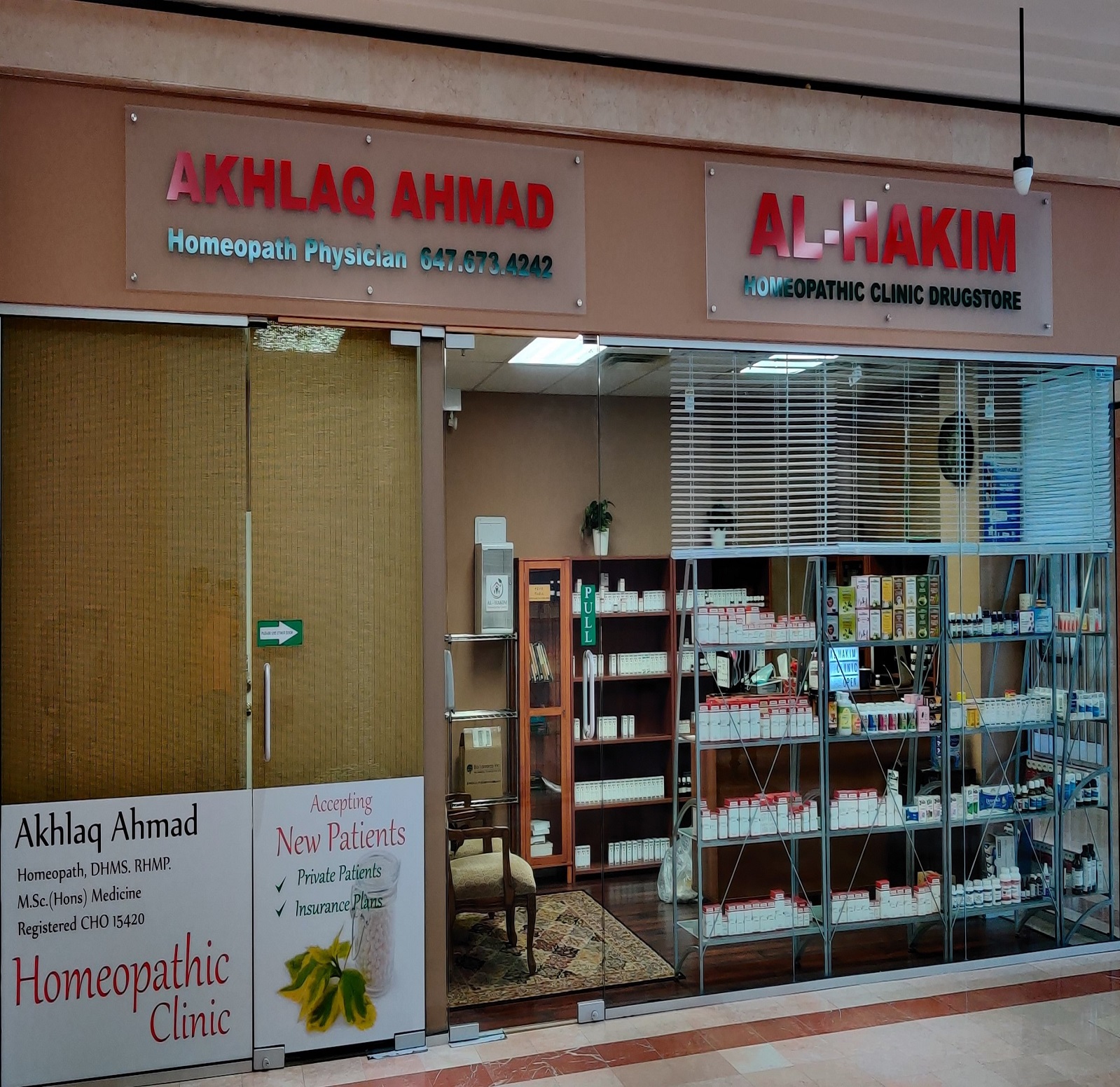
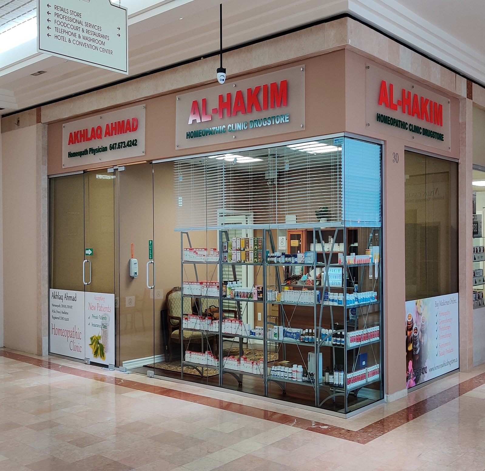
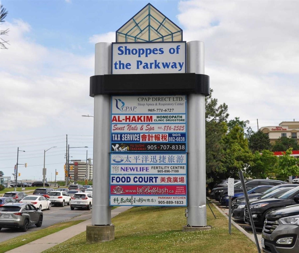
AL-HAKIM HOMEOPATHIC CLINIC
HOMEOPATH OFFICE
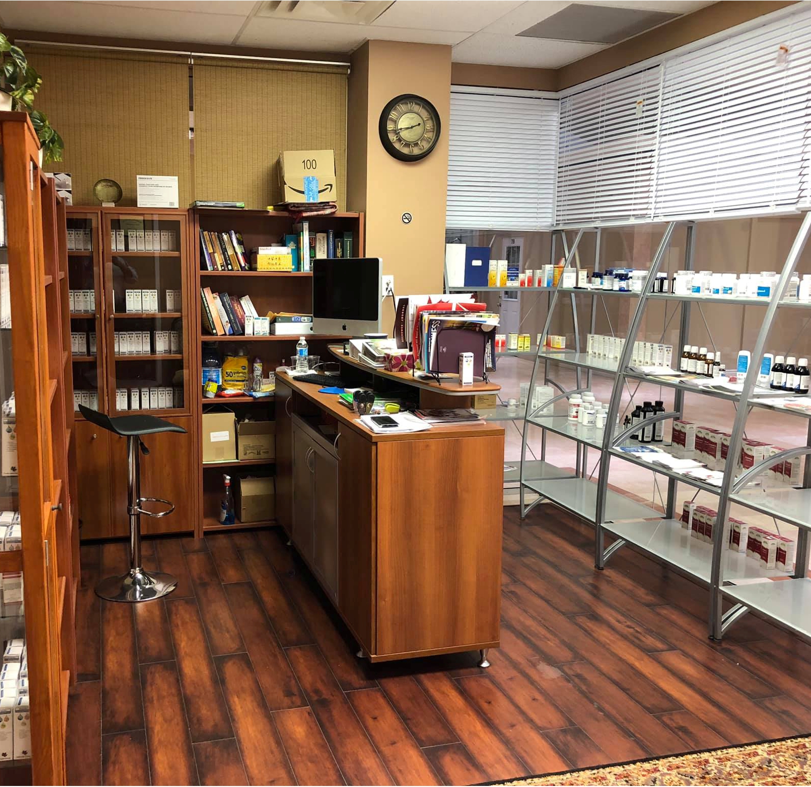
HOMEOPATHIC DRUGSTORE
BOOK AN APPOINTMENT HOMEOPATHIC CONSULTATION
Book Your Consultation Time in Advance while coming for your first Homeopathic Consultation Visit. Please bring with you all information/reports you may have from any other Health Care Professional you are seeing.
This website uses cookies so that we can provide you with the best user experience possible. Cookie information is stored in your browser and performs functions such as recognising you when you return to our website and helping our team to understand which sections of the website you find most interesting and useful.
Strictly Necessary Cookies
Strictly Necessary Cookie should be enabled at all times so that we can save your preferences for cookie settings.
If you disable this cookie, we will not be able to save your preferences. This means that every time you visit this website you will need to enable or disable cookies again.